Figure 3.
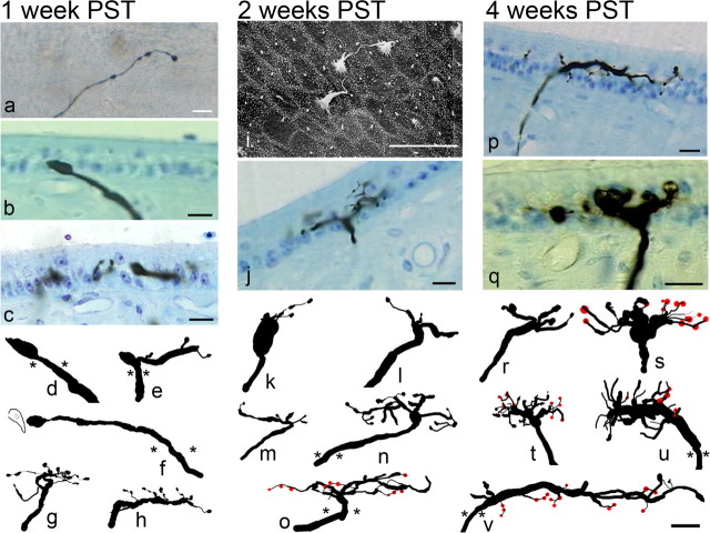
Photomicrographs and anatomical reconstructions of stage 1 regenerating afferents. a–h, Afferents innervating the maculas at 1 week PST were primarily composed of a simple initial fiber segment, prominent swellings, and a large terminal growth cone (a, surface view of saccular afferent; b, cross section of utricular afferent; d, f, reconstructions). Simple branched segments were also noted for some fibers, all ending in terminal growth cones (c, e, g, h). i–o, Photomicrographs and reconstructions for 2 week PST regenerated afferents. i, Scanning electron microscopic image of the apical surface of the central striola region in 2 week PST utricular macula. Morphological polarizations of stereocilia bundles were appropriately polarized toward the developing reversal line (as determined by epithelial location). j–o, Simple branched afferent structures with terminal growth cone processes directed toward extant hair cells were observed for most afferents. A few more complex branching patterns were also observed (n, o), and some segments contained bouton terminals (red circles). p–v, Afferents at 4 weeks PST exhibited more numerous short branched segments, most with growth cones. Some fiber segments contain boutons terminaux (p). In the saccular maculas, large rounded terminal growth cones were observed, many with branching segments emanating from prominent swellings (q). Reconstructions show that at 4 weeks PST, many fibers had multiple branches. A mixture of growth cones and bouton terminals was observed. Asterisks denote penetration point into the sensory epithelium. Scale bars, 10 μm.

