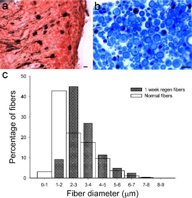
Figure 4.
Macular afferent number and fiber size after denervation of the epithelium (1 week PST). a, Vestibular ganglion with BDA-filled (black) and nonfilled neurons. b, Cross section of one utricular nerve branch immediately beneath the macula. BDA-filled (black) and nonpositive fiber lumens (blue) were measured. c, Histogram comparison of fiber diameters for normal (open; n = 2351) and 1 week PST fibers (hatched; n = 1831). Scale bars, 10 μm.

