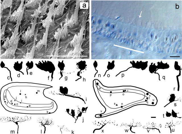
Figure 8.
Photomicrographs and anatomical reconstructions for stage 3 regenerated macular afferents at 24–36 weeks PST. a, Scanning electron microscopy of central striolar region in 24 week PST saccular macula. b, DIC photomicrograph of central striolar region in 24 week PST utricular macula. Eight type II cells in the type II band (white bar) flank the polarization reversal line (arrow). On either side of the band lie the medial (left) and lateral (right) striolar regions, with a dense concentration of type I hair cells (I) contained in calyceal terminals. c–u, Reconstructions for utricular (c–k) and saccular (n–u) afferents for stage 3 regeneration. Calyx afferents (c–f, n–q) arranged in increasing order of complexity. Calyx afferents were found primarily in the central regions. i, j, r–t, Dimorph afferents arranged in increasing order of complexity, with the distribution of the fibers in the regenerated maculas being more dispersed compared with normal development (see supplemental material, available at www.jneurosci.org). k–m, u–x, Reconstructions for seven regenerated bouton afferents arranged in increasing order of complexity. Insets, Composite surface maps of stage 3 regeneration utricular (left) and saccular (right) maculas. Distributions of reconstructed calyx (diamonds), dimorph (squares), and bouton (circles) afferents are shown. Scale bars, 10 μm.

