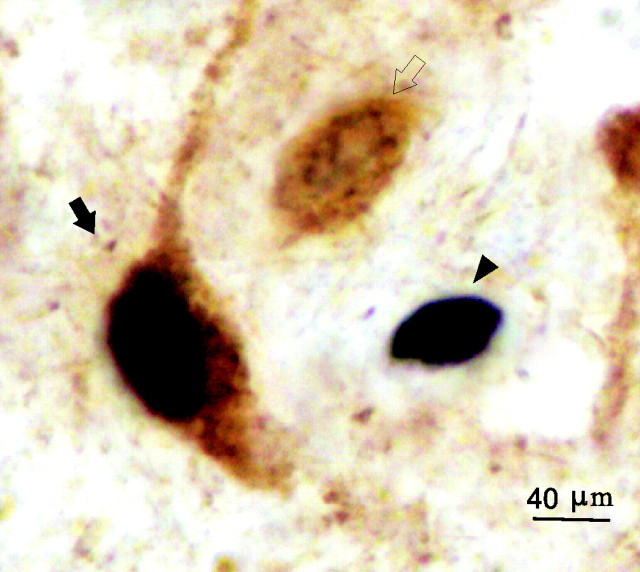
Figure 1.
Examples of Fos single-labeled (filled arrowhead), GAD single-labeled (open arrow), and Fos+ GAD double-labeled (filled arrow) neurons in the MnPN. The Fos protein is stained black and confined to the nucleus. The GAD-IRNs are stained brown, and the staining is evident throughout the soma and the proximal dendrites.

