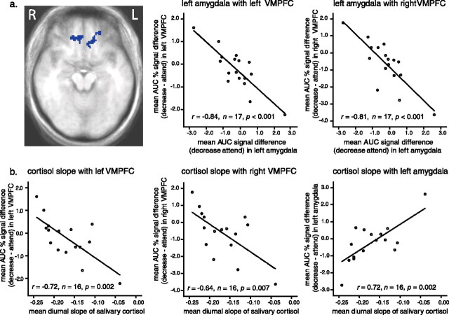
Figure 3.
a, Two regions in left (L) and right (R) VMPFC (maximum, t(16) = −4.79 at Talairach coordinates x = −23, y = 43, z = −10; and maximum, t(16) = −5.28 at x = 5, y = 37, z = −12, respectively) demonstrate an inverse across-subjects correlation with the left amygdala. For illustrative purposes, we extracted mean signal across all voxels for the decrease and attend conditions separately for the two VMPFC regions and also for the left amygdala. We then computed zero-order correlations between the amygdala and VMPFC across subjects and depicted those associations in the accompanying scatter plots. Subjects with lower activation in the amygdala (top middle and right) (abscissa) exhibit higher activation in the right and left VMPFC (ordinate). Represented on all axes is the difference in AUC for percentage signal change between the decrease and attend conditions (decrease–attend). b, We next computed zero-order, across-subjects correlations between diurnal cortisol slope (abscissa) and the difference in AUC for percentage signal change between the decrease and attend conditions (decrease–attend) across all voxels in the left and right VMPFC (bottom left and middle) and the amygdala (bottom right) (ordinate) clusters. Subjects with higher activation in VMPFC and lower activation in the amygdala exhibit the steepest declines in salivary cortisol over the course of the day.

