
Figure 4.
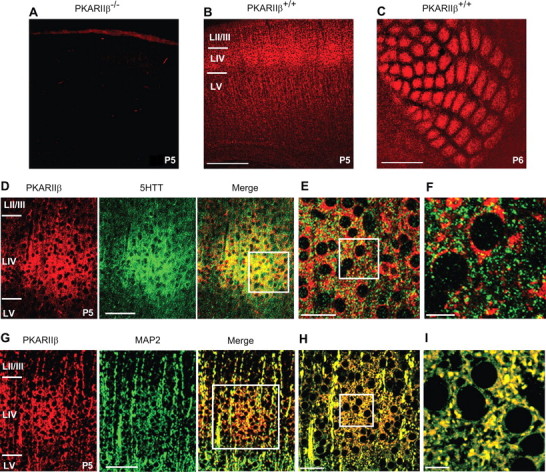
PKARIIβ is extensively colocalized with postsynaptic but not presynaptic markers. A, Control immunostaining of a P5 PKARIIβ−/− thalamocortical slice with anti-PKARIIβ antibody reveals that this antibody is specific to PKARIIβ. B, C, Cortical expression of PKARIIβ was examined by labeling wild-type thalamocortical (B) and tangential (C) slices with anti-PKARIIβ antibody. PKARIIβ is expressed in layer IV as it delaminates from layer II/III at P3 (data not shown) and forms a barrel pattern when barrels emerge at around P5 (B, C). D–I, Presynaptic and postsynaptic localization of PKARIIβ was examined by coimmunostaining P5 wild-type thalamocortical slices using anti-5-HTT (D–F) and anti-MAP2 (G–I) antibodies, respectively, together with an anti-PKARIIβ antibody. D, Low-magnification image of a single barrel stained with a 5-HTT antibody (green) to label presynaptic thalamocortical terminals and a PKARIIβ (red) antibody and their merged image. At low magnification, both show a clear barrel pattern. E, Merged image of the barrel in D (white box) at 63× magnification. F, A 4× zoom of white-boxed area in E. G, Low-magnification image of a single barrel labeled with a MAP2 antibody (green) to mark postsynaptic dendrites and a PKARIIβ antibody (red) and their merged image. H, Merged image of the barrel in G (white box) at 63× magnification. I, 4× zoom of white-boxed area in H. Colocalization analysis of high-magnification images using ImageJ shows that PKARIIβ expression colocalizes significantly more (p < 0.05; t test) with MAP2 (28.8 ± 7.5%; n = 4) than with 5-HTT (7.1 ± 2.2%; n = 4). LIV, Layer IV; LV, layer V. Scale bars: A–C, 500 μm; D, G, 100 μm; E, H, 40 μm; F, I, 10 μm.