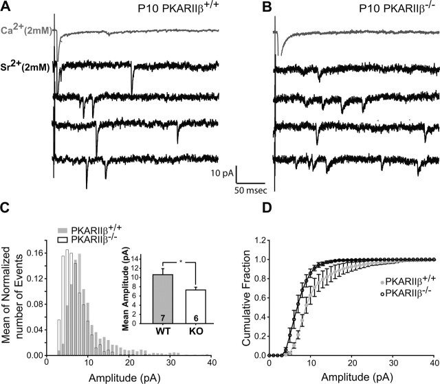
Figure 6.
Small AMPAR mini-EPSCs at PKARIIβ−/− thalamocortical synapses. A, B, Sample traces of evoked AMPAR-mediated current responses in Ca2+–ACSF (gray) and Sr2+–ACSF (black) from recordings in P9–P11 PKARIIβ−/− (B) and wild-type littermate control (A) neurons. In the presence of Sr2+–ACSF, responses to evoked synchronous release is lower and quantal events (evoked mini-EPSCs) in response to asynchronous release appear. The amplitudes of these quantal events are smaller on average in PKARIIβ−/− mice. C, Average frequency histogram of evoked mini-EPSCs. The PKARIIβ−/− histogram (white bars) peaks at a smaller amplitude than wild-type littermate controls (gray bars), which have a longer large amplitude tail. Inset, Mean evoked mini-EPSC amplitude is significantly lower (*p < 0.05; t test) for PKARIIβ−/− mice (white) with respect to wild-type littermate controls (gray). D, The cumulative probability distribution in PKARIIβ−/− (KO) animals (black) also shows a shift toward smaller amplitudes compared with wild-type (WT) littermate controls (gray). Comparison of root mean square noise shows no difference (p = 0.45; t test) between genotypes (data not shown). Error bars indicate SEM.

