
Figure 3.
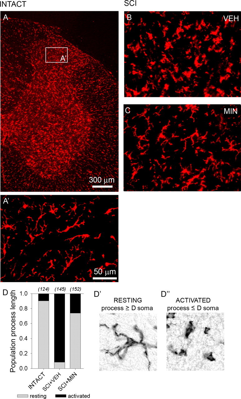
Immunolabeling for OX-42-positive microglia. OX-42 signal revealed moderate expression of resident microglia in both white and gray matter of the lumbar dorsal horn in an intact spinal cord (A). Microglia exhibited the resting type morphology: small compact somata bearing long, thin, ramified processes (A’). Thirty-three days after SCI, in vehicle-treated animals (SCI+VEH), microglia exhibited the activated phenotype: marked cellular hypertrophy and retraction of processes (B). Very few cells exhibiting resting morphological features were detected. In SCI animals, on day 33, after 3 d of i.t. administration of minocycline (SCI+MIN), microglia assumed the resting morphology (C). Quantification (D) of the proportion of resting (D’) and activated (D”) microglia confirmed that after SCI, there is a large shift from resting to activated microglia and that minocycline significantly reversed this shift.