
Figure 8.
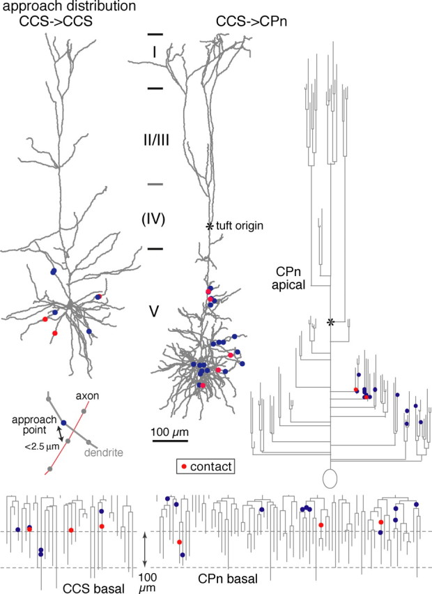
Distributions of approach points along reconstructed dendrites and dendrograms of example postsynaptic neurons (basal dendrites of a CCS cell, and basal and apical dendrites of a CPn cell). Contact sites are shown in red, and noncontact approach points are shown in blue. Inset, Example of an approach point (blue circle) of an axon (red line) to a dendrite (gray line). Reconstructed axons and dendrites were composed of serial points (circles) with intervals shorter than 1.5 μm. Approach points were defined as dendritic sites where axons passed within 2.5 μm.