
Figure 5.
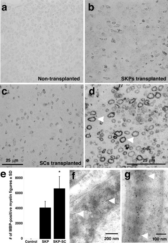
Structural evidence of myelination in the injured peripheral shiverer nerve by SKP-derived Schwann cells. Either naive SKPs or purified, SKP-derived Schwann cells were transplanted distal to a sciatic nerve crush in immunosuppressed shiverer mice, and nerves were analyzed as semithin cross sections 4 weeks after transplantation. a–c, Photomicrographs of semithin cross sections immunostained for MBP and visualized with DAB, showing the contralateral, uninjured, nontransplanted shiverer nerve (a), a nerve transplanted with naive SKPs (b), or a nerve transplanted with SKP-derived Schwann cells (c). Note the relatively robust number of MBP-positive myelin figures in the transplanted but not contralateral nerves. d, Higher-magnification micrograph illustrating the morphology of the MBP-positive myelin figures (arrowheads). Similar results were obtained in five transplanted nerves in each group. e, Quantitation of MBP-positive myelin figures in cross sections similar to those shown in a–d, comparing the relative number of myelin figures in nerves transplanted with naive SKPs versus SKP-derived Schwann cells. Details about the method of relative quantitation are provided in Results. Error bars indicate mean ± SD. ∗p < 0.05 for the comparison between the naive SKPs and SKP-derived Schwann cells, ANOVA; n = 3 in each group. f, g, Electron micrographs of ultrathin cross sections of injured shiverer sciatic nerve 4 weeks after transplantation of purified SKP-derived Schwann cells. Sections were immunostained for MBP and visualized with gold-labeled secondary antibody. Note the gold particles on the compact myelin (arrowheads).