
Figure 1.
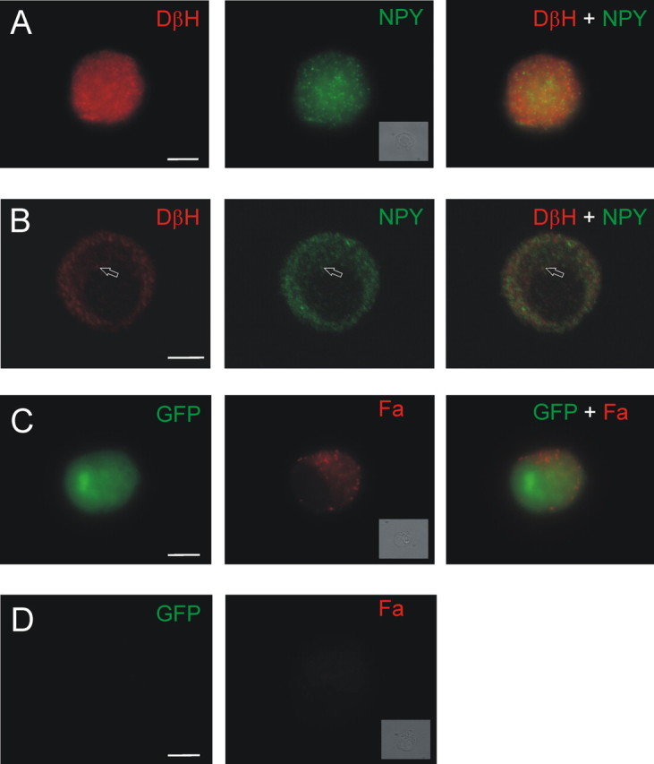
Markers of catecholamines and peptides are found in the same chromaffin cells. A, Example of a chromaffin cell that was double stained with antibodies for DβH (left) and NPY (middle). Right, Merged image. B, Single confocal slice indicates that the NPY and DβH immunoreactivity is punctate and occasionally colocalized (arrow). C, Chromaffin cell cotransfected with GFP and FMRFamide (Fa)-tagged NPY prohormone. GFP (left) fills the cell, whereas FMRFamide immunoreactivity (middle) is localized to puncta. Right, Merged image. D, A nontransfected, GFP-negative cell (left) is not FMRFamide immunoreactive (right; see Materials and Methods). A, C, D, Insets, Bright-field images of each cell. Scale bars, 5 μm.