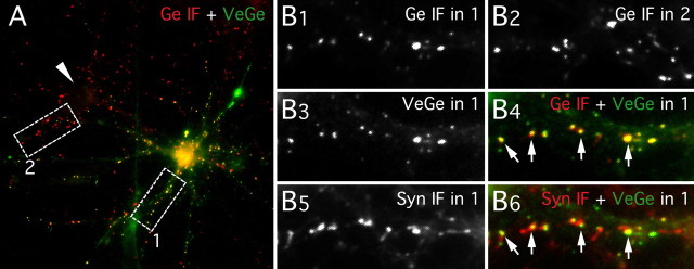
Figure 1.
Synaptic localization of Venus::gephyrin. Synapsin I (Syn) and gephyrin (Ge) IF labeling in a spinal cord neuron culture (10 DIV) transfected with VeGe is shown. A, Gephyrin-immunopositive clusters (red) in a transfected neuron (YFP fluorescence, green) and in a nontransfected neuron (arrowhead). B, Higher magnification of areas 1 and 2 (white boxes in A) showing gephyrin-immunopositive clusters in area 1 (B1) and area 2 (B2), VeGe cluster intrinsic fluorescence in area 1 (B3), Syn I-positive presynaptic terminals in area 1 (B5), and superimposition of VeGe fluorescence (green) and gephyrin IF (red; B4) or synapsin I IF (red; B6) puncta in area 1. Note (1) that Venus::gephyrin (A, B3) and endogenous gephyrin clusters (A and B2) display similar distribution patterns and comparable sizes in a transfected neuron and in a nontransfected neuron, respectively; (2) that all gephyrin IF clusters apposed to presynaptic terminals (arrows in B4 and B6) contain VeGe molecules; and (3) most VeGe clusters are apposed to presynaptic terminals (B6).

