
Figure 3.
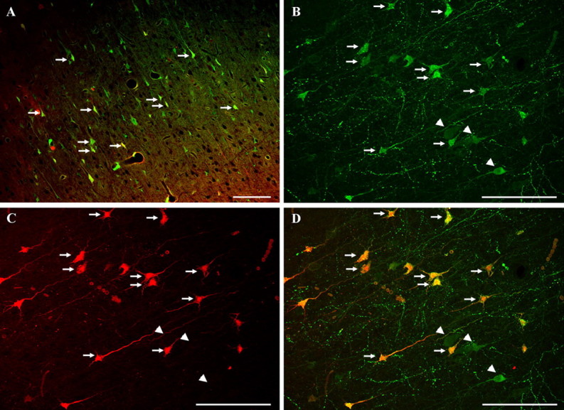
Confocal images of tracer-infused-injured animals. A, This double-labeled image demonstrates numerous cortical neurons (arrows) revealing intracellular tracer flooding with both the preinjury and postinjury infused dextrans. Scale bar, 100 μm. B–D, These confocal images, revealing the initially administered dextran flooding (B), the postinjury infused dextran flooding (C), and their overlay (D), demonstrate that the majority of the neurons sustaining membrane disruption and tracer uptake are double labeled both with preinjury- and postinjury-infused dextrans (arrows). Such double labeling was observed at both 4 and 8 h after injury. Note that some neurons flooding with the preinjury dextran alone can also be observed (arrowheads). Scale bar, 100 μm.