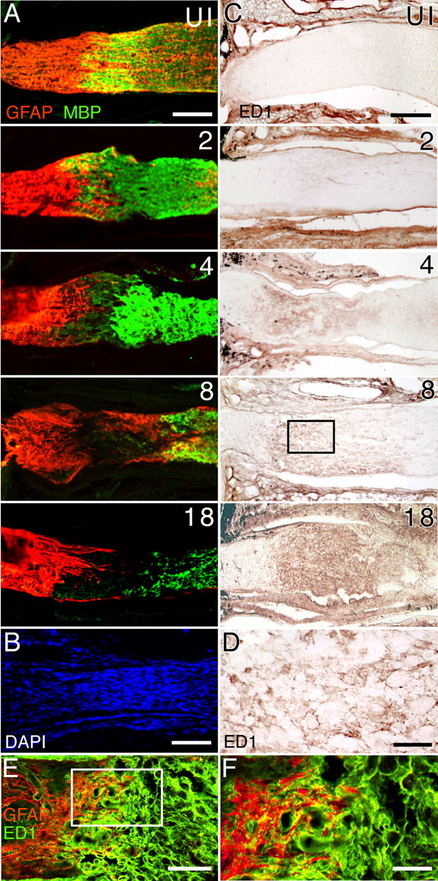
Figure 2.

Cellular reorganization in the optic nerve after injury. A, Distribution of GFAP+ astrocytes (red) and myelin basic protein (MBP+) oligodendrocytes (green) before and after nerve injury. The numbers represent the days after injury. Retina is toward the left. By 4 d after injury, astrocytes and oligodendrocytes have begun to vacate the injured area, and a region devoid of these two cell types is observed by 8 d after injury. B, DAPI cell nuclei staining from a different optic nerve showing the presence of numerous cell bodies at the lesion site 12 d after injury. C, Accumulation of ED1+ macrophages after optic nerve injury. The boxed region at day 8 after injury is shown at a higher magnification in D. D, Higher-magnification view of ED1+ macrophages. E, Double immunostaining of an optic nerve tissue section 8 d after injury using anti-GFAP (red) and anti-ED1 (green) showing that the margin region surrounding the lesion site contains both astrocytes and macrophages. F, Higher-magnification view of the boxed region in E. Scale bars: A–C, E, 100 μm; D, F, 20 μm. UI, Uninjured nerve.
