Figure 4.
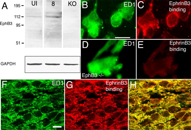
Expression of EphB3 protein in the injured adult optic nerve and macrophages. A, Immunoblots of protein preparations from uninjured (UI) and injured adult optic nerves 8 d after injury (8) probed using an anti-EphB3 antibody. A signal at ∼110 kDa corresponding to the expected size for EphB3 protein was detected in both samples. This protein band was not present in optic nerve tissue obtained from EphB3 null animals (KO). GAPDH-immunopositive bands used as protein loading controls are shown below. B, ED1-positive macrophages isolated from segments of injured adult optic nerve in culture. C, The macrophages shown in B bound exogenously applied EphrinB3-Fc protein resulting in a punctate–aggregate pattern of cell-surface labeling. D, ED1-positive macrophages from segments of injured adult optic nerve in culture derived from an EphB3 homozygous null animal. E, Exogenously applied EphrinB3-Fc protein failed to bind to the EphB3 null macrophages shown in D. F, ED1-positive macrophages in an adult optic nerve 5 d after injury (green). G, EphrinB3-Fc binding in the same region shown in D (red). H, Colocalization of ED1 immunoreactivity with EphrinB3-Fc binding. Scale bars: (in B) B–E, 20 μm; (in F) F–H, 10 μm.

