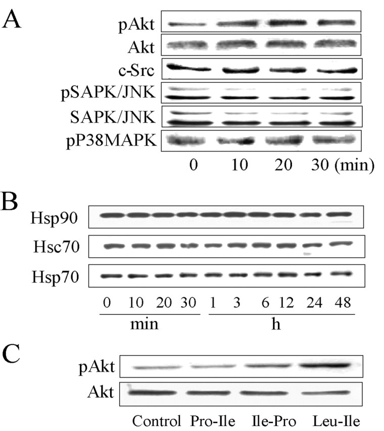
Figure 4.

Leu-Ile stimulates Akt phosphorylation. A, Neurons were exposed to Leu-Ile (10 μg/ml) for the indicated times. Cell lysates were subjected to SDS-PAGE and probed with various antibodies. The representative immunoblots are shown. B, Neurons were exposed to Leu-Ile (10 μg/ml) for the indicated times. Immunoblots were probed with antibodies against Hsp90, Hsp70, and Hsc70. C, Neurons were stimulated with Leu-Ile, Pro-Leu, and Ile-Pro (10 μg/ml) for 30 min. Cell lysates were subjected to SDS-PAGE and probed with antibodies against pAkt and Akt.
