
Figure 5.
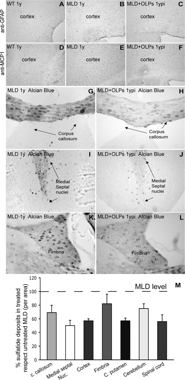
Sulfatide deposits and gliosis were reduced in MLD mice grafted with OLPs 1 year after surgery. Decreased level of astrogliosis (GFAP) and microgliosis (MCP-1) was observed in treated animals (C, F) in comparison to 1-year-old untreated MLD (B and E, respectively). A, D, Endogenous expression of GFAP and MCP-1 in healthy wild-type brain. Sections of transplanted (H–L) and nontransplanted (G–K) MLD brains were stained with Alcian blue to detect sulfatide deposits. Corpus callosum (H), medial septal nuclei (J), and the fimbria (L) of transplanted MLD mice showed reduction in the number and size of sulfatide deposits in comparison to the untreated MLD brain (G, I, K). M, The graph shows the abundance of sulfatide deposits in different areas of the brain expressed as a percentage of deposits in the same areas from untreated mice. n = 5 mice per group. ypi, Year postinjection; c. callosum, corpus callosum; Medial septal Nuc., medial septal nuclei; C. putamen, caudate–putamen. Results are expressed as mean value ± SEM.