
Figure 3.
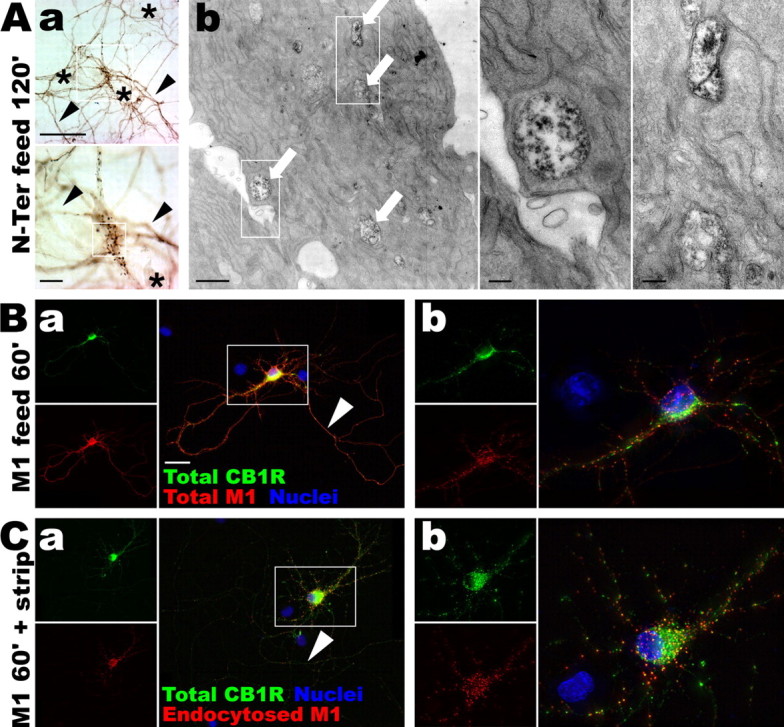
CB1R is constitutively endocytosed, preferentially in the somatodendritic compartment. A, Living neurons at DIV 9 are fed for 2 h with the N-Ter antibody directed against the extracellular N terminus of the endogenous CB1R. Aa, After permeabilization and immunoperoxidase staining, the N-Ter antibody labels axons (arrowheads) and somatodendritic vesicles in the soma of CB1R-positive neurons (boxed). Neurons not expressing CB1R do not uptake the N-Ter antibody (asterisks). Scale bars: top, 50 μm; bottom, 10 μm. Ab, Correlated electron microscopy of the same neuron showing labeled endosomes within the soma (arrows) that have endocytosed the N-Ter antibody. Scale bars: left, 1 μm; right panels, 0.2 μm. B, DIV 9 neurons expressing FCB1-eGFP (green) are fed for 1 h with anti-FLAG M1 antibody (red). The M1 antibody labels axons (Ba, arrowhead) and shows punctate pattern of CB1 red labeling on the soma and dendrites (Bb), indicating constitutive endocytosis of CB1R. Scale bar, 20 μm. C, Neurons expressing FCB1-eGFP are fed for 1 h with M1 antibody. Surface-bound antibodies are then stripped by an acid wash, leaving only primary antibodies bound to endocytosed receptors. After fixation and permeabilization, red secondary labeling shows few endocytosed receptors in the axon (Ca, arrowhead) whereas the somatodendritic compartment contains numerous antibody-labeled endosomes (Cb), showing that constitutive endocytosis of CB1R occurs primarily in the somatodendritic compartment.