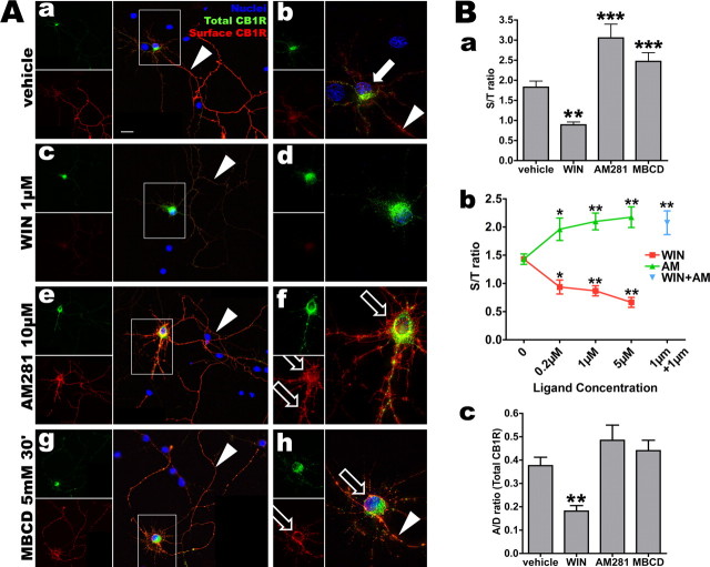
Figure 5.
Somatodendritic CB1R trafficking is dependent on the pharmacological state of CB1R. A, Neurons expressing FCB1-eYFP are incubated for 3 h with vehicle (0.2% DMSO; Aa, Ab), WIN (1 μm; Ac, Ad), AM281 (10 μm; Ae, Af), or 30 min with MBCD (5 mm; Ag, Ah), then rapidly labeled for surface receptors with M1 antibody and fixed. eYFP signal (in green) corresponds to total CB1R population and surface labeling is revealed in red. Left panels (Aa, Ac, Ae, Ag) show the neuron with its axonal arborization (montage of two images) and right panels (Ab, Ad, Af, Ah) are a projection of a deconvoluted 10 μm z-stack (0.2 μm z-step) taken with a 100× objective. Images for vehicle, WIN, and AM281 conditions were acquired and processed identically to preserve relative staining intensities. Scale bar: (in Aa) Aa–Ah, 20 μm. In control cells, surface CB1R is found on the axonal arborization (Aa, arrowhead) whereas somatodendritic receptors are mostly endosomal (arrow in Ab, highlighting the soma of the transfected neuron). Note the axon on Ab that is brightly labeled for surface CB1R just after the axon hillock (arrowhead). After WIN treatment, somatodendritic surface receptors are barely visible, whereas endosomes are abundant in the soma and dendrites (Ac, Ad). Axonal surface receptors are downregulated but still present (Ac, arrowhead). Somatodendritic surface labeling is strongly enhanced after AM281 treatment (Af, empty arrows) and axonal surface labeling is still strong (Ae, arrowhead). Blocking endocytosis with MBCD leads to a CB1R distribution similar to AM281 treatment with upregulation of surface expression of CB1R on the somatodendritic plasma membrane (Ah, empty arrows). B, Quantifications of CB1R translocation in neurons transfected with FCB1-eYFP (corresponding to images shown in A). Ba, Transfected neurons are quantified using the S/T ratio of 24–36 neurons per condition from two to three independent experiments. WIN downregulates surface population of CB1Rs leading to a drop of the S/T ratio, whereas inverse agonist AM281 upregulates plasma membrane receptors, similarly to endocytosis blocker MBCD, that lead to a rise of the S/T ratio. Bb, Effects of both WIN and AM281 on CB1R localization are concentration-dependent (red curve for WIN, green curve for AM281) and resulting S/T ratios are significantly different from control for all concentrations tested. AM281 antagonizes WIN because coapplication of both ligands at 1 μm leads to upregulation of the surface receptor similarly to treatment with AM281 alone (blue data point). Bc, CB1R localization in transfected neurons is quantified as the A/D ratio, that is the ratio of intensities between dendrites and axon for total CB1R (green channel) in individual neurons in each condition (n = 24–26 neurons from two independent experiments). WIN is able to translocate CB1R from axon to soma, as shown by the decrease in A/D ratio, whereas AM281 and MBCD do not have a significant effect on the compartmentalization of CB1R between axonal and somatodendritic compartment. In all quantifications, values are mean ± SEM and asterisks show significance between vehicle and treated conditions, as defined in Materials and Methods.

