
Figure 2.
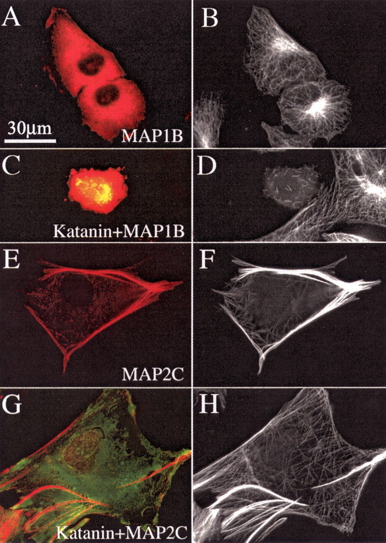
MAP2c protects microtubules from being severed by P60-katanin, but MAP1b does not. A, C, E, G, GFP–P60-katanin in green and immunostain for either plasmid-expressed MAP2 or MAP1b in red. B, D, F, H, Immunostains for microtubules. A, B, Cells that are not overexpressing P60-katanin. As shown in A and B, MAP1b expression does not cause abnormal bundling of microtubules. As shown in C and D, a cell overexpressing MAP1b and P60-katanin shows only a scattering of very short microtubules and severely reduced microtubule levels. (A neighboring cell not expressing MAP1b or P60-katanin displays a normal microtubule array). As shown in E and F, MAP2c expression causes the formation of dense bundles of microtubules. As shown in G and H, the microtubules in MAP2c-expressing cells show no indication of severing by overexpression of P60-katanin, and the microtubule mass is not reduced. Scale bar: A–H, 30 μm.