
Figure 7.
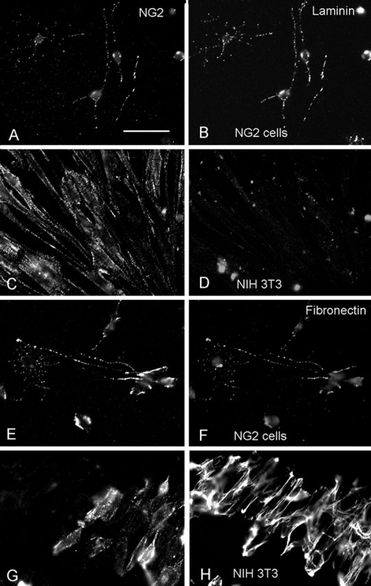
Cell surface expression of laminin and fibronectin on NG2 cells. Double-immunofluorescence labeling for NG2 (A, C, E, G; detected with Alexa 594 goat anti-rabbit immunoglobulins) and laminin (B, D) or fibronectin (F, H), detected with Alexa 488 goat anti-mouse immunoglobulins, on secondary cultures of NG2 cells from P3 rat cortex (A, B, E, F) and mouse NIH 3T3 cell fibroblasts (C, D, G, H). Scale bar: (in A) A–H, 24.4 μm. Both laminin and fibronectin are colocalized with NG2-immunoreactive puncta on rat NG2 cells. Little laminin is detected on NIH 3T3 cells. Fibronectin on NIH 3T3 appears extracellular.