Figure 2.
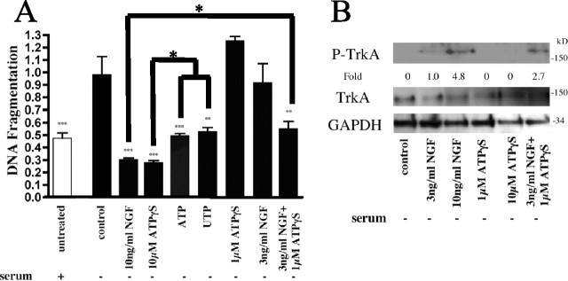
ATPγS/NGF interact to enhance inhibition of apoptosis. Serum-starved PC12 cells were treated with ATP (100 μm), UTP (100 μm), NGF, and/or ATPγS at the indicated concentrations. Apoptosis was quantitated using DNA fragmentation (A). An immunoblot analysis of cells treated with NGF (10 or 3 ng/ml), ATPγS (10 or 1 μm), or 3 ng/ml NGF together with 1 μm ATPγS and probed for TrkA activation is shown (B). Densitometry was measured as P-TrkA/TrkA and normalized to 3 ng/ml NGF treatment alone. *p < 0.05, **p < 0.01, ***p < 0.001 versus serum starvation alone, with values normalized to serum starvation alone. Error bars indicate SE.

