
Figure 7.
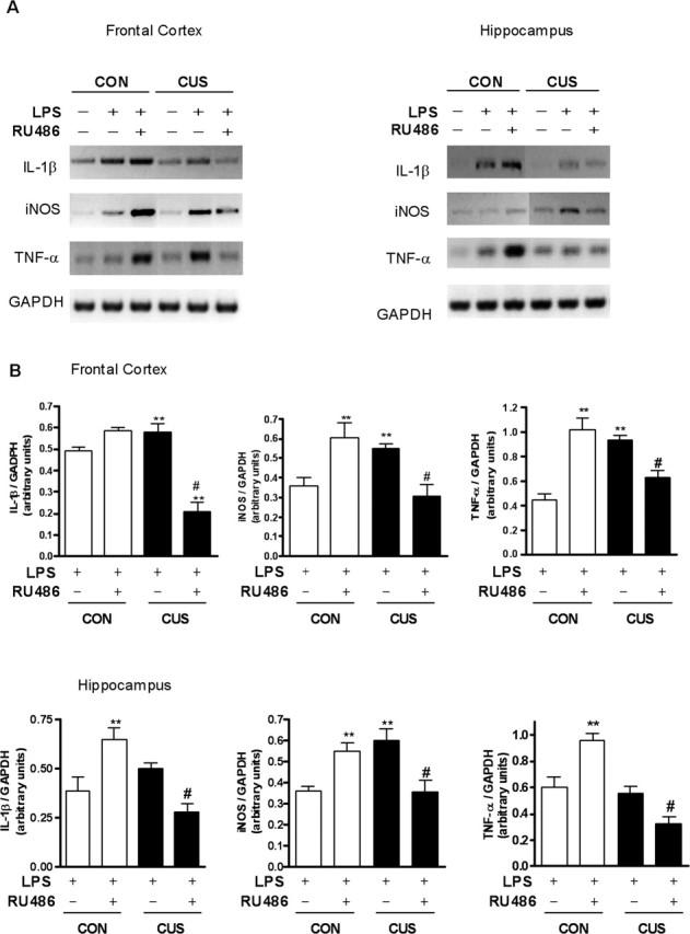
Influence of RU-486 on mRNA levels of proinflammatory genes (IL-1β, iNOS, and TNF-α) induced by LPS in the frontal cortex and hippocampus of unstressed (CON) and stressed (CUS) rats. A, Representatives PCR photographies. B, Desitometric analysis of the specific bands of LPS-treated groups presented in A. mRNA levels is presented as ratios of target gene to GAPDH expression. Data are presented as mean ± SEM; n = 4 animals per group. Bonferroni’s test: **p < 0.05 versus CON plus LPS; #p < 0.05 versus CUS plus LPS.