
Figure 1.
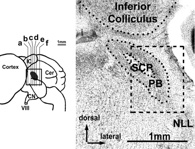
Reconstruction of the location of PB (gray) in a side view of the horseshoe bat brain (left) and histograph of frontal section through PB (right). Letters in the left graph indicate positions of sections shown in Figure 2A-F. The histograph shown here corresponds to c. Box represents the total area covered by the reconstruction shown in Figure 2 and depicts the relative position of the PB within the area covered in Figure 2C. VIII, Auditory nerve; Cer, cerebellum; CN, cochlear nucleus; IC, inferior colliculus; NLL, nuclei of the lateral lemniscus; SCP, superior cerebellar peduncle.