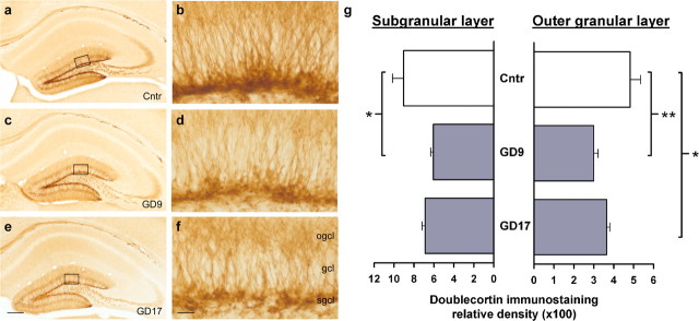
Figure 4.
Reduction in postnatal neurogenesis in the dentate gyrus after prenatal immune challenge. a, c, e, Photomicrographs of coronal brain sections of the hippocampal formation and dentate gyrus were taken from representative control offspring (a) and animals subjected to prenatal PolyI:C exposure on GD9 (c) or GD17 (e) and visualized with immunoperoxidase staining using anti-DCX antibody. b, d, f, As evident in the images at a higher magnification (indicated by the squares in a, c, and e), reduced DXC relative optical density was observed in both the outer granule cell layer and the subgranular cell layer of the dentate gyrus after prenatal PolyI:C exposure, and this effect was primarily independent of prenatal treatment times. The values in g are mean ± SEM. ∗p < 0.05 and ∗∗p < 0.01, statistical significance based on Fisher's LSD post hoc analysis. Scale bars: a, c, e, 500 μm; b, d, f, 50 μm. Cntr, Control; ogcl, outer granule cell layer; gcl, granule cell layer; sgcl, subgranular cell layer.

