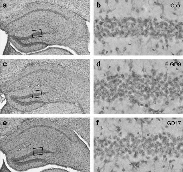
Figure 6.
Unaltered postnatal brain gross morphology after prenatal PolyI:C exposure. a, c, e, Photomicrographs of coronal brain sections of the dorsal hippocampal formation were taken from representative control animals (a) and animals subjected to prenatal PolyI:C exposure on GD9 (c) or GD17 (e) and visualized with Nissl/GFAP double staining, which stains neuronal cell bodies and astrocytes, respectively. b, d, f, Note that images on higher magnification (indicated by the squares in a, c, and e) revealed no differences between prenatal PolyI:C-treated [GD9 (d) and GD17 (f)] and control (Cntr; b) offspring in the neuronal cell numbers or in the abundance of pyknotic neurons and astrocytes. Scale bars: a, c, e, 500 μm; b, d, f, 50 μm.

