
Figure 2.
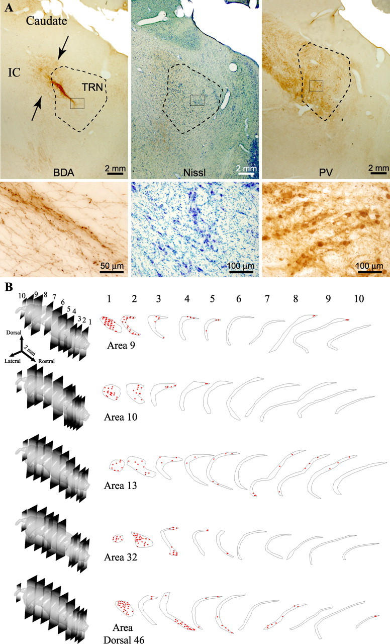
Prefrontal axonal terminations in the reticular nucleus. A, Corticothalamic pathway from area 32 to TRN (arrows in left column), labeled with BDA, entering through the internal capsule (IC), shown at low magnification (top) and high magnification (bottom). The middle and right columns show matched sections at low (top) and high (bottom) magnification stained with Nissl (middle) or immunostained for PV (right) to show the borders of TRN. B, Two-dimensional quantitative projection patterns in 10 representative coronal sections covering the whole rostrocaudal extent of TRN, shown in 3D reconstructions on the left. Each set of outlines corresponds to axonal boutons (red dots) from one prefrontal area. Each dot represents ∼10 boutons.