
Figure 2.
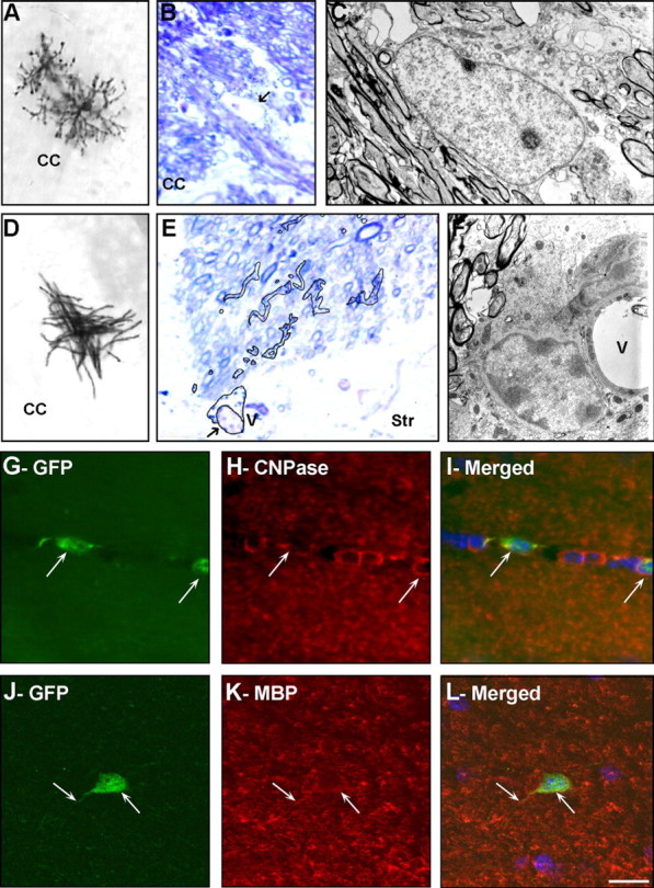
SVZ astrocytes give rise to oligodendrocytes in the adult mouse brain. RCAS–AP (A–F) or RCAS–GFP (G–L) retroviruses were injected into the SVZ of adult Gtv-a mice, and the progeny of the infected SVZ astrocytes was analyzed 21 d after injection using light (A, B, D, E), confocal (G–L), and electron (C, F) microscopy. A–F, Several AP-labeled cells were identified in the CC (A–D) and in the striatum (E, F): highly branched cells presenting typical morphology of nonmyelinating (A–C) and myelinating (D–F) oligodendrocytes. Characteristics of AP+ nonmyelinating oligodendrocytes (B, C) and AP+ mature myelinating oligodendrocytes (E, F) were determined at the ultrastructural level. B, E, AP-staining in 1.5-μm-thick semithin sections counterstained with toluidine blue. Arrows indicate the nucleus of the cells analyzed at the electron microscopic level. In E, AP+ cell body and processes were traced on top of the picture. C, F, Electron micrograph of AP+ cells. Note the different ultrastructure and typical nuclear heterochromatin in myelinating oligodendrocytes compared with that of the nonmyelinating one. G–L, Double immunofluorescence for GFP (G, J) and the oligodendrocyte markers CNPase (H) or MBP (K) of cells in the CC. Merged field shows combined immunostaining for GFP and CNPase (I) or MBP (L). Arrows indicated SVZ-generated cells in the CC-expressing markers of mature oligodendrocytes. Str, Striatum; V, blood vessel. Scale bar: A, D, 80 μm; B, E, 7 μm; C, F, 2 μm; G–L, 20 μm.