
Figure 1.
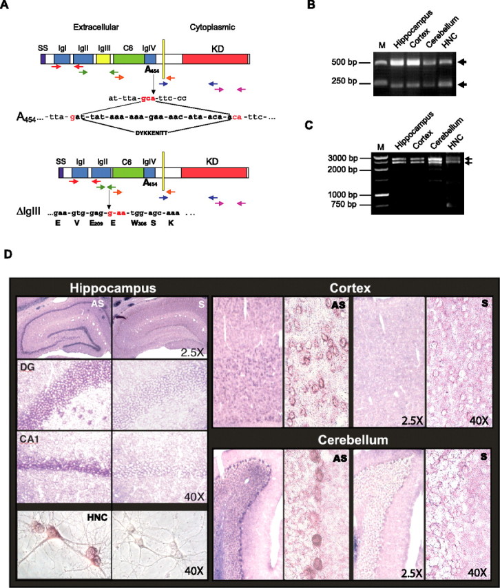
MuSK is expressed in adult rat brain. A–C, Cloning of MuSK in the brain. A, Schematic representation of MuSK isoforms expressed in the brain. SS, Signal sequence; IgI–IgIV, Ig-like domains; C6, cysteine rich domain; KD, kinase domain; E, glutamic acid; W, tryptophan; S, serine; V, valine; K, lysine. Arrows in different colors indicate the five sets of primers used for PCRs. B, Electrophoretic analysis of PCR amplifications generated with the second set of primers (shown in green in Fig. 1A). M, Molecular weight marker. C, Electrophoretic analysis of PCR amplifications with a set of primers that flanked the entire MuSK ORF. D, Musk mRNA distribution in adult rat brain and HNCs revealed by in situ hybridization. Representative examples of rat adult brain coronal sections and HNCs hybridized with antisense (AS) or sense (S) probes. DG, Dentate gyrus. CA1 regions of the hippocampus, somatosensory cortex, cerebellum, and HNCs are shown. Magnifications: 2.5 and 40×. Data are representative of results obtained from eight experiments.