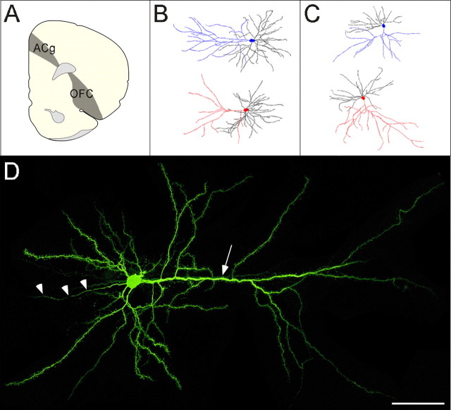
Figure 1.
A, Coronal hemisection of the prefrontal cortex [bregma 3.20 mm, adapted from Swanson (1992)] depicting ACg and lateral OFC regions of interest. B, C, Dendritic reconstructions of neurons from ACg (B) and lateral OFC (C), with apical dendrites highlighted in blue (controls) and red (stressed). D, A typical pyramidal neuron from lateral OFC, with the apical dendrite (arrow) extending from the soma toward the pial surface at right and the axon (arrowheads) extending to the left. Scale bar, 50 μm.

