
Figure 1.
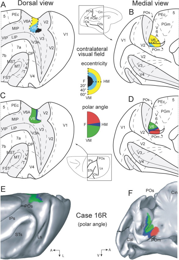
Topology and visual topography of macaque monkey area V6. A–D, Dorsal (A, C) and medial (B, D) views of the left hemisphere of a macaque monkey brain (modified from Galletti et al., 1999a). PO, POM, inferior occipital (IOS), occipitotemporal (OTS), intraparietal (IPS), lunate (LS) and superior temporal (STS) sulci are opened to reveal areas in the depth of these sulci. Opened sulci are depicted in light gray, and are indicated as thickened lines on the brain silhouettes at the center of the figure. Dashed lines are the borders between different cortical areas, according to Colby et al. (1988), Desimone and Ungerleider (1986), Galletti et al. (1999a,b), Gattass et al. (1981, 1988), Gattass and Gross (1981), and Pandya and Seltzer (1982). In A and B, the map of eccentricity in V6 is color-coded: central, parafoveal, and peripheral are represented as black, cyan, and yellow, respectively. C, D, The polar angle map of V6 is reported; lower, horizontal, and upper fields are represented as green, blue, and red, respectively. E, F, Dorsal and medial views of surface reconstructions of case 16R (Galletti et al., 1999a) performed using CARET software (see Materials and Methods). Polar angles of receptive fields recorded from V6 are indicated using the same color scale as in C and D. HM, Horizontal meridian; VM, vertical meridian; DP, dorsal prelunate; VIP, ventral intraparietal; LIP, lateral intraparietal; MST, medial superior temporal; FST, fundus of the superior temporal; MIP, medial intraparietal.