
Figure 6.
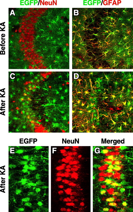
Hippocampal CA3 neurons express EGFP, representing apoE, in response to excitotoxic injury. Heterozygous 5-month-old EGFPapoE mice received peritoneal injections of kainic acid (KA; 25 mg/kg) (C–G), and the brains were collected 1 d (E–G) or 6 d (C, D) later. Untreated age-matched heterozygous EGFPapoE mice served as controls (A, B). Confocal images of immunostained brain sections were collected for EGFP (green) and anti-NeuN (red), a neuronal marker, or anti-GFAP (red), an astrocytic marker. Images in A–D and G are merged, and yellow indicates colocalization.