
Figure 3.
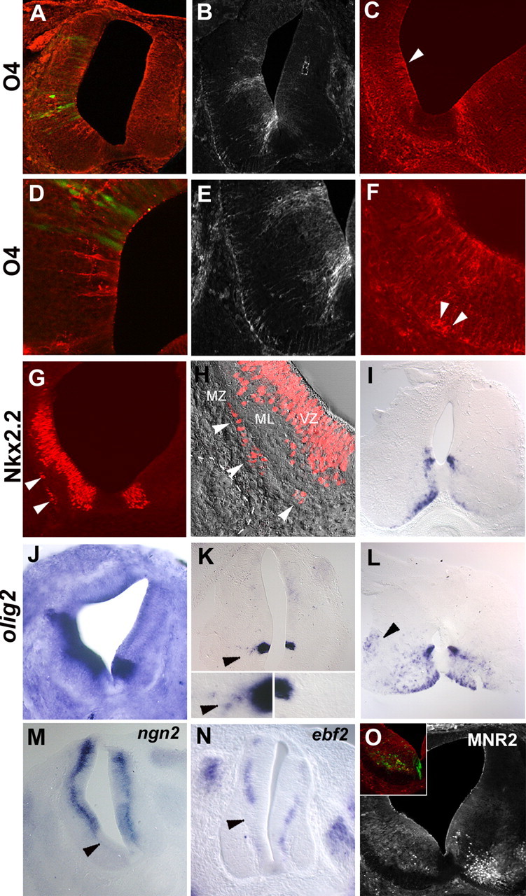
Elevating the doses of Shh in the E1.5 neural tube induces premature OLP specification and inhibits neuronal production in the ventral spinal. Early neural progenitors (E1.5) have been submitted to high activity of Shh by electroporation of a Shh-expressing vector (A–F, I, K–M, O) or graft of Shh-expressing cells (G, H, J, N). The side of electroporation or graft is represented on the left in each picture. A, B, D, E, Electroporation of a Shh-expressing vector results at E3.5 in the emergence of premature O4-expressing cells (red) in the ventral spinal cord (A, D). At E4.5, numerous prematurely induced O4+ cells were detected (B, E). Note the absence of O4 immunoreactivity in the control side at both stages. D, E, High magnifications showing that O4+ cells are located in the neuroepithelium. C–F, At E6, electroporation of Shh causes dorsal extension of the OL domain (C, arrowhead) compared with the nonelectroporated side (C, right). At higher magnification, O4+ cell bodies can be already detected at the periphery of the spinal cord (F, arrowheads). G–I, Graft of Shh-expressing cells results at E4 in dorsal extension of the Nkx2.2-expressing domain of the ventricular zone. Note the presence of ectopic Nkx2.2+ cells located in the marginal zone (G, arrowheads), whereas such cells are not detected in the contralateral side. MZ, Marginal zone; ML, mantle layer; VZ, ventricular zone. H, Overlay of immunostaining and Nomarski images at high magnification of ventrolateral part of grafted spinal cord showing the presence of Nkx2.2+ cells in the forming marginal zone (white arrowheads delineate the pial surface of the spinal cord; the dashed line indicates the position of grafted QT6-Shh cells). At E8, numerous nkx2.2-expressing cells were accumulated in the marginal zone, whereas only a few of them were detected in the control side (I). J–L, Detection of olig2 transcript by in situ hybridization. At E4, a marked extension of the olig2-expressing domain is observed in grafted embryo (J). At E4.5/E5 in an electroporated embryo, few olig2+ cells emigrating from the Olig2-expressing neuroepithelial domain were observed (K, arrowheads), whereas such migration is not noticed in the control side (K). The insets in K show high magnification of olig2-expressing domains of the ventral neuroepithelium and olig2+ emigrating cells in the electroporated side (arrowhead). At E8, olig2-expressing cells have already reached the intermediate marginal zone of the spinal cord (L, arrowhead). M–O, Inhibition of ventral neuronal genesis by high doses of Shh in the embryonic spinal cord. Detection of neuronal precursors using ngn2 (M) and ebf2 (N) on transverse sections of E4 embryos electroporated or grafted at E1.5. Note the extinction of neuronal markers on the operated side (M, N, arrowheads) compared with the contralateral side. O, At E6, immunodetection using MNR2 shows a strong inhibition of MN differentiation in the electroporated side (O, inset).