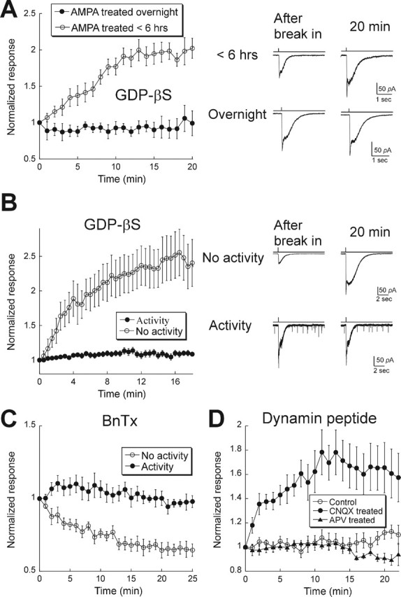
Figure 2.

AMPAR trafficking rate is decreased by excitatory activity. A, Left, Cultured retinal neurons were incubated with 10 μm AMPA and 50 μm APV overnight (filled symbols; n = 5) or for <6 h (range, 1–5 h; open symbols; n = 11). Right, Examples of responses after break-in (left) and 20 min later under each condition. B, Retinal–hippocampal cocultures. Left, Summary of results for cells with (filled symbols; n = 19) or without (open symbols; n = 13) sEPSCs. All cells were dialyzed with GDP-βS. Right, Records taken from two cells, one with and the other without excitatory synaptic activity. Records were obtained at break-in and 20 min later. Note the presence of sEPSCs in the bottom panel. C, Summary of results from cells in retinal–hippocampal cocultures dialyzed with BnTx. In those cells with spontaneous EPSCs, BnTx had no effect, as expected in the absence of rapid AMPAR cycling (filled symbols; n = 5). Conversely, in cells without spontaneous EPSCs, BnTx produced a time-dependent depression of the response to AMPA puffs (open symbols; n = 5). D, Mixed retinal–hippocampal cultures were incubated overnight in media alone (control; open circles; n = 19) or media containing 50 μm CNQX (filled circles; n = 11) or 50 μm APV (filled triangles; n = 7). Inhibition of AMPARs, but not NMDARs, was sufficient to restore rapid AMPAR cycling. Error bars represent SEM.
