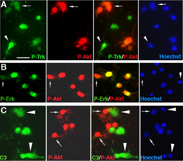
Figure 9.
Pro-NGF induces apoptosis of BDNF-treated BF neurons with activated Trk receptors but not with phosphorylated Akt or Erk. A, Neurons were double immunostained for P-Trk and P-Akt and labeled with Hoechst. Anti-P-Akt labeled a subset of the P-Trk-positive neurons that were not apoptotic (arrows). The arrowheads indicate apoptotic neurons that are P-Trk positive and P-Akt negative. B, Neurons were double labeled for P-Akt and P-Erk. P-Erk was present in most but not all of the P-Akt-positive population, which were all healthy. An arrow indicates a P-Akt-positive neuron that lacks P-Erk. An arrowhead indicates an apoptotic cell that had neither P-Akt nor P-Erk. C, Neurons were double labeled for P-Akt and cleaved caspase-3 (C3) and labeled with Hoechst. The arrows indicate healthy neurons, and the arrowheads indicate apoptotic neurons. All of the cleaved caspase-3-positive neurons were apoptotic and lacked P-Akt, whereas all of the P-Akt-positive neurons were healthy and lacked cleaved caspase-3. Scale bar: (in A) A–C, 20 μm.

