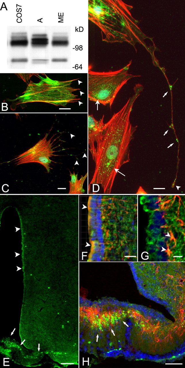
Figure 2.

The TACE protein is present in hypothalamic glial cells and in the intact rat hypothalamus. A, Immunoblotting of Sepharose–concanavalin A-enriched cell lysates derived from hypothalamic astrocytes (A), the ME of the hypothalamus, and COS-7 cells (positive control) demonstrates the presence of a major immunoreactive species migrating at ∼110 kDa under the nonreducing conditions used and that represent the full-length, unprocessed TACE. A smaller species of ∼75–80 kDa corresponding to TACE lacking the prodomain (Schlöndorff et al., 2000) was also detected in the ME and COS cells and to a lesser extent in astrocytes. Protein samples were boiled in sample buffer in the absence of dithiothreitol and size fractionated on a 8–16% polyacrylamide gel before immunoblotting (see Material and Methods). B, Detection of astrocytic processes not stained by GFAP antibodies, using phallotoxin conjugated to Texas Red. GFAP staining is shown as a green color; arrowheads point to some of the processes devoid of GFAP. C, D, Localization of TACE in cultured hypothalamic astrocytes. TACE was identified with rabbit polyclonal antibodies to its cytoplasmic tail domain, and the reaction was developed to a green color with Alexa 488 streptavidin. Thereafter, the cells were stained with phallotoxin conjugated to Texas Red to visualize actin in astrocytic processes that are devoid of GFAP staining. Notice that TACE immunoreactivity is present at the tip of astrocytic processes (C, arrowheads), in addition to having a perinuclear localization (D, long arrows). TACE is also detected along fine processes in which it appears to be most abundant at the tip of the processes (D, arrowhead) and at bifurcation points (D, short arrows). E–H, Detection of TACE immunoreactivity in the hypothalamus of immature 30- to 32-d-old rats. Astrocytes were identified as such with monoclonal antibodies to GFAP. Notice that, in this case, the immunoreaction was developed to a red color using Texas Red goat anti-mouse IgG. E, TACE immunoreactivity (green) is present in cells scattered throughout the hypothalamus, but it is particularly evident in ependymoglial cells (tanycytes) lining the third ventricle (3V; arrowheads) and the ME (arrows). F, Three-dimensional rendering of a stack of images of the periventricular region showing the presence of TACE in ependymoglial cells (arrowheads) identified as tanycytes by vimentin immunostaining (red). Notice that TACE is also present in ependymoglial processes. G, In addition to ependymoglial cells (green), TACE is also present in subependymal astrocytes (arrowheads). H, TACE (green) is abundant in astrocytes (red) of the ME (arrows), but a substantial fraction of the TACE-immunoreactive material appear not to overlap with the GFAP staining, indicating the presence of TACE in GFAP-negative astrocytic processes. Blue, Hoechst nuclear stain. Scale bars: B, D, F,20 μm; C,10 μm; E, 100 μm; G,15 μm; H,40 μm.
