
Figure 2.
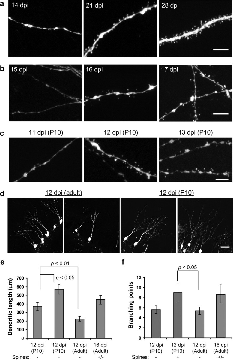
The initiation of spine genesis in newly generated granule neurons. a, Examples of dendritic segments from mouse brains taken at 14, 21, and 28 dpi. Scale bar, 5 μm. b, Examples of dendritic segments at 15, 16, and 17 dpi. Scale bar, 5 μm. c, Examples of dendritic segments of neurons born at P10. Spines start to grow before 12 dpi (middle panel). Scale bar, 5 μm. d, Representative images of more mature-looking cells at 12 dpi. Scale bar, 50μm. e, Quantification of dendritic length of cells born in the adult brain at 12 and 16 dpi, as well as cells born at P10 at 12 dpi. f, Quantification of branching points. Data are presented as mean ± SEM in e and f.