Figure 1.
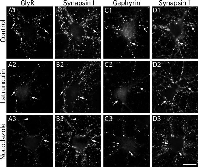
Effects of F-actin and microtubule disruption on GlyR and gephyrin clusters. Double detection of GlyR (A) and synapsin (B) or gephyrin (C) and synapsin (D) in cultured spinal cord neurons (10–12 DIV). GlyR (A1–A3) and gephyrin (C1–C3) formed aggregates (arrows) apposed to presynaptic terminals (B1–B3 and D1–D3, respectively) in control conditions (A1–D1) or after 1 h treatment with 3 μm latrunculin (A2–D2) or 10 μm nocodazole (A3–D3). Note that GlyR-, gephyrin-, but not synapsin-associated immunoreactivity decreased in the presence of latrunculin or nocodazole. Scale bar, 20 μm.

