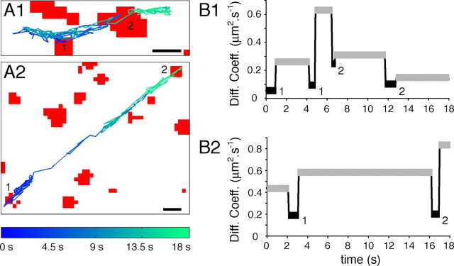
Figure 4.
Decrease in GlyR diffusion coefficients at synapses independently of cytoskeleton-disrupting treatments. A, Reconstruction of 18 s GlyR-QD mass center trajectory over the FM4-64-stained synapses (red) images after 1 h treatment with latrunculin (A1) or nocodazole (A2). Scale bars, 1 μm. B, Comparison of diffusion coefficients averaged during extrasynaptic (gray) and synaptic (black) sequences from the trajectory illustrated in A in latrunculin (B1) or nocodazole (B2) condition. Numbers correspond to synapses in A. Note that, despite the latrunculin- and nocodazole-induced increase in GlyR lateral mobility, receptors are consistently slowed down at synapses.

