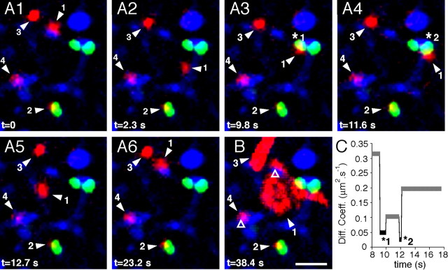
Figure 5.
GlyR lateral diffusion at inhibitory synapses. Example of a recording session of GlyR-QDs on neurons transfected with Vege chimera in the presence of latrunculin. Blue, FM4-64-stained synapses; red, GlyR-QDs; green, Vege-containing inhibitory postsynaptic microdomains. A1–A6, Sequence of pictures extracted from a 38.4 s recording session at the indicated times. B, Maximum intensity projection showing the surface area explored by GlyR-QDs during the recording session. Various diffusive behaviors are found in this field. GlyR-QD 1 swaps between a Vege-devoid synapse (B, open triangle) and Vege-containing synapses (A3, star 1; A4, star 2). GlyR-QD 2 remains at the same Vege cluster. GlyR-QD 3 and 4 are mobile in the extrasynaptic membrane and stable at a Vege-devoid synapse (B, open triangle), respectively. Scale bar, 2 μm. C, Averaged diffusion coefficients of GlyR-QD 1 during extrasynaptic (gray) and synaptic (black) sequences.

