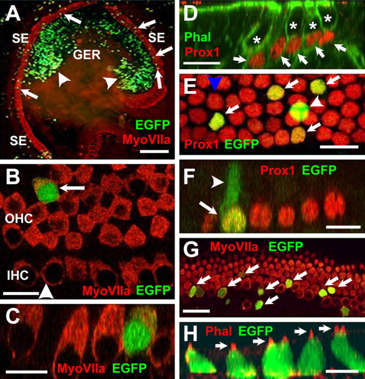
Figure 4.

Transfection of progenitor cells in cochlear explant cultures. A, Low-magnification view of an explant culture electroporated at E13 with an EGFP expression plasmid and maintained in vitro for 6 d. Hair cells within the sensory epithelium (SE) have been labeled with anti-myosin VIIa (red), and transfected cells expressing EGFP are green. EGFP-positive cells (green) are present in the GER (arrowheads) and the SE (arrows). B, High-magnification view of the sensory epithelium in an EGFP-transfected explant. The single row in inner hair cells (IHC) and, in this case, four rows of outer hair cells (OHC) are positive for myosin VIIa (red). A single transfected cell (green) has developed as an outer hair cell (arrow). The arrowhead indicates the plane of Z-section in C. C, Confocal Z-stack cross section along the plane indicated by the arrowhead in B, illustrating hair cell morphology in the transfected cell (green). D, Prox1 is a marker for supporting cells in the organ of Corti. Confocal Z-stack of a P0 mouse cochlea. Prox1 expression is illustrated in red. Filamentous actin is labeled with phalloidin in green. Nuclei of supporting cells (arrows) are positive for Prox1 (red), whereas nuclei of hair cells (asterisks) are negative for Prox1. E, High-magnification view of the supporting cell nuclear layer in an explant transfected with EGFP. Four transfected cells (green, arrows) are also positive for Prox1. A single additional cell (arrowhead) is also transfected but appears to be located at a more lumenal position within the epithelium and is not Prox1 positive. This cell has probably developed as a hair cell. The blue arrowhead indicates plane of Z-section in F. F, Confocal Z-stack cross section through the plane indicated by the blue arrowhead in E. The single transfected Prox1-positive cell (arrow) has a morphology that is consistent with a supporting cell, including an apical projection (arrowhead) that extends to the lumenal surface. G, Overexpression of Math 1 induces development of hair cells in the sensory epithelium. Virtually all progenitor cells transfected with Math 1 (green, arrows) are positive for myosin VIIa (red), indicating that they have developed as hair cells. H, Math 1-transfected cells also develop stereociliary bundles. Confocal Z-stack cross section through a group of transfected progenitor cells (green). Stereociliary bundles on each hair cell (arrows) are visualized by phalloidin labeling of filamentous actin (red). Scale bars: A, 250 μm; B, 20 μm; C, 10 μm; D, 20 μm; E, 20 μm; F, 20 μm; G, 20 μm; H, 10 μm.
