Figure 2.
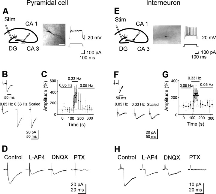
Minimal stimulation of granule cells in the dentate gyrus evokes GABAA-mediated monosynaptic responses in CA3 pyramidal cells and interneurons. A, E, Left, Diagrams of the hippocampus showing a CA3 pyramidal cell (A) and an interneuron (E) with converging inputs from MFs. Stim, Stimulating electrode. Middle, Neurons labeled with biocytin. Scalebar, 50 μm. Right, Representative traces showing the typical firing pattern of a CA3 pyramidal cell and an interneuron at P3 in response to long depolarizing current pulses. B, F, Paired stimuli delivered at 50 ms interval to granule cells in the dentate gyrus evoked at -70 mV synaptic responses exhibiting strong paired-pulse facilitation (average of 50 individual responses). Bottom, Mean amplitude of synaptic responses (average of 15 individual traces) evoked by stimulation of granule cells in the dentate gyrus at 0.05 and 0.33 Hz. On the right, the two responses are normalized and superimposed. C, G, The mean amplitude of synaptic currents evoked in three pyramidal cells (C) and in three interneurons (G) at 0.05 and 0.33 Hz (bars) is plotted against time. Note the slow buildup of facilitation of synaptic responses at 0.33 Hz that completely reversed to control values after returning to 0.05 Hz stimulation. Error bars represent SEM. D, H, Synaptic responses from another pyramidal cell and interneuron before and during application of l-AP-4 (10 μm) and l-AP-4 plus DNQX (20 μm). Addition of picrotoxin (PTX; 100 μm) completely abolished synaptic currents. Each response is the average of 20 individual traces (including failures). Note that GABAA-mediated synaptic currents were depressed by l-AP-4 but were unaffected by DNQX.

