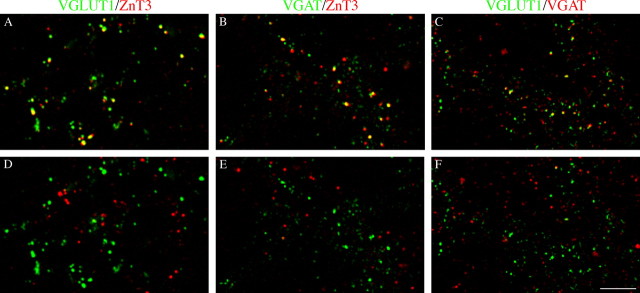
Figure 9.
Immunocytochemical evidence for the expression of VGAT in MF terminals. VGAT expression in CA3 VGLUT1- and ZnT3-positive fibers at P5. A, VGLUT1/ZnT3-positive puncta. B, C, VGLUT1- and ZnT3-positive puncta coexpress VGAT immunoreactivity in PL. D-F, Rotation of the red channel by 90° strongly reduces colocalization. SL, Stratum lucidum. Scale bar: (in F) A-F, 5 μm.

