
Figure 2.
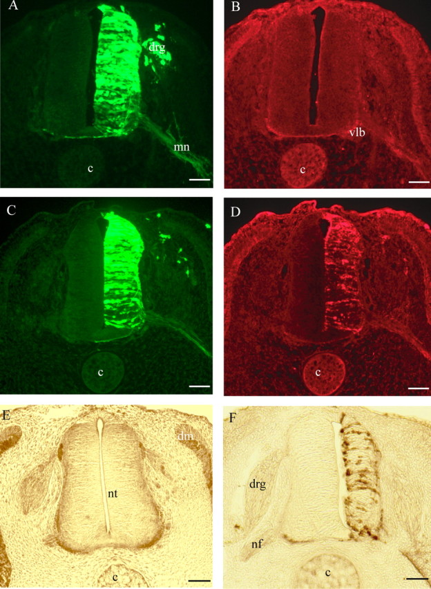
Cross sections of electroporated neural tubes from chick embryos. A–D, HH stage 21 embryos electroporated with the following pCAGGS expression vectors: GFP (A, B) and GFP plus Alix (C, D). A, C, GFP 24 h after electroporation in the neural tissue; GFP is also visible in dorsal root ganglia (drg) and in the axons from motoneurons (mn). Vlb, Ventrolateral border; c, cord. B, D, Immunofluorescence revealing endogenous and overexpressed Alix, respectively, by using polyclonal anti-Alix antibody recognizing endogenous chick and overexpressed mouse Alix. Scale bars, 50 μm. E, F, HH stage 24 embryos, 48 h after electroporation, immunostained with polyclonal anti-Alix revealed by peroxidase. E, Control embryo transfected with GFP pCAGGS plasmid alone. Endogenous Alix is strongly expressed ventrally and laterally along the border of the neural tube (nt). Alix is also expressed in the nerve fibers (nf) and in the dermomyotome (dm). F, Alix-transfected embryo. The neuroepithelium of the electroporated side is clearly reduced in size. Revelation was shorter in F compared with E to avoid saturation of the overexpressed protein and reducing background. Scale bars, 50 μm.