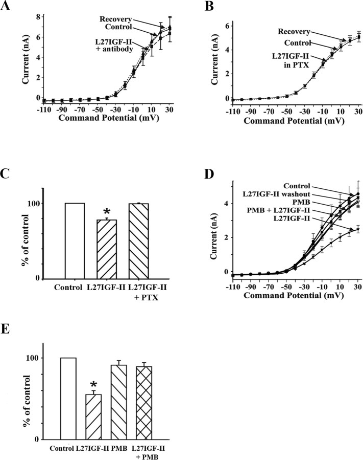
Figure 8.
A, I-V relationship from DBB neurons (n = 9) pretreated with IGF-II/M6P receptor antibody (1:100), which showed a drastically reduced response to Leu27IGF-II (L27IGF-II; 50 nm) in whole-cell currents. B, I-V relationship from DBB neurons (n = 12) pretreated with PTX (1 μg/ml), in which Leu27IGF-II (50 nm) did not evoke a significant reduction in whole-cell currents. C, Histograms show the effects of Leu27IGF-II alone (n = 17) and in the cells pretreated with PTX (n = 12) as a percentage of control whole-cell currents at +30 mV; *p < 0.01. D, I-V relationship from DBB neurons (n = 7) in which Leu27IGF-II evoked a significant and reversible reduction in whole-cell currents. In the same cells, inclusion of PMB (10 μm) in the perfusion medium blocks the Leu27IGF-II-induced reduction of whole-cell currents. E, Histograms depict the effects of Leu27IGF-II, PMB, and Leu27IGF-II in the presence of PMB (n = 7) as a percentage of control whole-cell currents at +30 mV; *p < 0.01.

