
Figure 1.
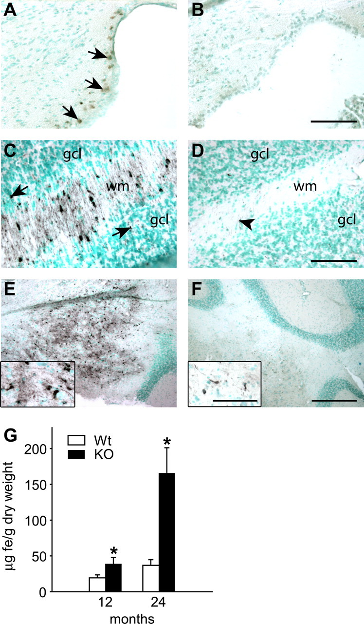
Iron accumulation in the cerebellum of Cp−/− and Cp+/+ mice with age detected by enhanced Perl's histochemistry. Tissue sections are counterstained with methyl green. A, B, Twelve-month-old cerebellum. C–F, Twenty-four-month-old cerebellum. A, The region adjacent to the lining of the fourth ventricle shows the first evidence of iron accumulation (arrows) seen as brown staining in Cp−/− mouse at 12 months of age. B, No iron accumulation is detected in a wild-type mouse at this age. C, The white matter (wm) of the cerebellar cortex of a Cp−/− mouse shows marked accumulation of iron in the glial cells. Some iron-positive cells (arrows) are also seen in the granular cell layer (gcl). D, A few iron-positive cells (arrowhead) are seen in the cortical white matter (wm) and granule cell layer (gcl) in a wild-type mouse. E, Low-magnification micrograph of the deep nuclei of a Cp−/− mouse shows severe iron accumulation. F, A small number of iron-positive cells are seen in the deep nuclei of a wild-type mouse. Insets show higher magnification of some of the iron-containing cells. Scale bars: A–D, 100 μm; E, F, 400 μm; insets, 50 μm. G, Changes in iron concentration in cerebellum of Cp−/− knock-out (KO) and Cp+/+ wild-type (Wt) mice obtained by fAAS analysis. Data are presented as average ± SD. *p < 0.05; n = 3.