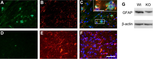
Figure 7.
Double-immunofluorescence labeling of cerebellar white matter at 24 months of age with anti-ferritin and anti-GFAP antibodies. A, B, Tissue section through the white matter of a Cp−/− mouse shows numerous ferritin-positive cells (A) that are double labeled with anti-GFAP (B), indicating that they are astrocytes. C shows merged images of ferritin, GFAP, and DAPI staining. The boxed area in C is shown at a higher magnification in the inset. D, An occasional ferritin-positive cell is seen in the white matter of wild-type mouse. B, E, Note that the extent and intensity of the GFAP labeling is markedly diminished in the Cp−/− mouse compared with the wild type. F shows merged images of ferritin, GFAP, and DAPI staining. Scale bars: F, 50 μm; inset in C, 20 μm. G, Western blot shows a reduction in GFAP in Cp−/− knock-out (KO) mouse compared with control wild-type (Wt) mouse at 24 months of age.

