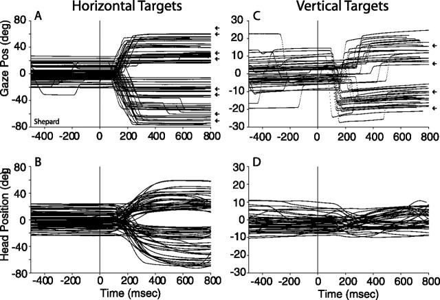
Figure 4.
Gaze and head movements to acoustic targets on the main horizontal (A, B) and vertical (C, D) axes recorded with the fixation task from subject Shepard. All data are plotted synchronized to the onset of the acoustic target at times 0 ms. A, C, The location of the targets is illustrated by small arrows plotted to the right of the gaze plots. The stimuli were 500–1000 ms broadband noise bursts. The secondary components of the gaze shifts and head movements were omitted for simplicity.

