Figure 2.

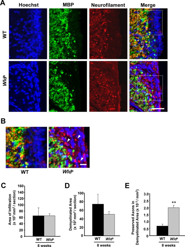
Wlds expression reduces axon loss at 8 weeks p.i. A, Representative images of transverse sections from EAE-induced wild-type (top panels) and Wlds (bottom panels) mice at 8 weeks p.i. stained with Hoechst 33258 (left), anti-MBP (middle), or anti-NF (right). B, The demyelinated areas are enlarged and the arrowheads indicate preserved axons in the demyelinated area. Scale bars: A, 30 μm; B, 10 μm. C–E, Quantification of the average areas of infiltration (p = 0.99) (C) and demyelination (p = 0.39) (D) per section, which showed no significant difference, and the average numbers of NF+/MBP− fibers in demyelinated areas, which was significantly increased in Wlds mice when compared with wild type (**p < 0.001; Student's t test) (E). Areas of infiltration (×103 μm2/section) or demyelination (×103 μm2/section) were determined as the measured Hoescht+ or MBP− areas averaged from 10 transverse sections per animal for each group. The number of preserved axons in demyelinated areas (×10−3/μm2) was determined by counting the number of NF+ puncta in demyelinated MBP− regions, and then dividing by the area of demyelination. Error bars indicate SEM.
