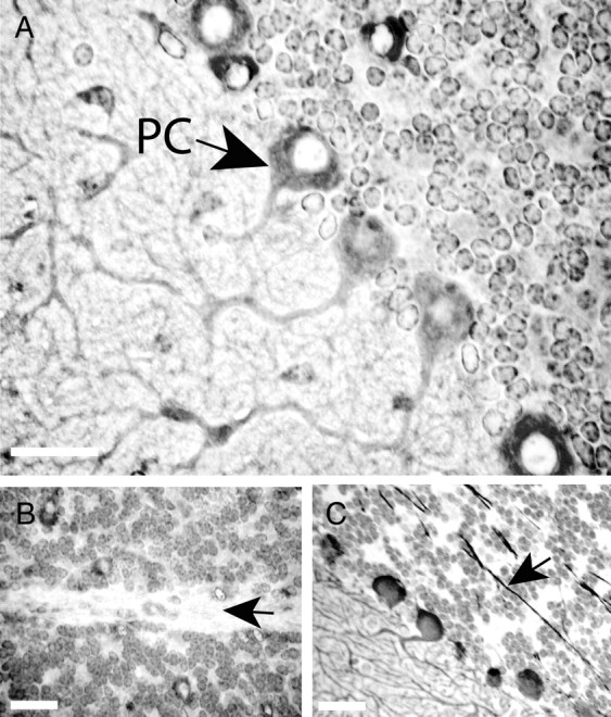
Figure 1.

Cellular and subcellular localization of Klhl1 in mouse cerebellar tissue. Immunohistochemical staining of normal FVB mice at 4 weeks of age with KLHL1-specific monoclonal antibody 13D9 shows that Klhl1 is a prominent cytoplasmic protein in the soma and dendrites of cerebellar Purkinje cells but is not present in the nuclei (A) or the axons (B) of these cells. Both the axons and the nuclei of Purkinje cells stain positively with anti-calbindin antibody (C). PC, Purkinje cell. Black arrows show the axons of the Purkinje cells. Scale bars, 25 μm.
